
SCIENCE
BENEFITS OF MICROSURFACING
Microsurfaced ADM (MS ADM) consists of human dermis that has been decellularized, disinfected and then processed using a proprietary microsurfacing method to create a multitude of thin, partial thickness cuts across the surface of the tissue. The resulting graft represents an advanced allograft solution that is designed to rapidly achieve desired clinical outcomes¹˒².
Key advantages vs. standard grafts include:
Significant increase in surface area at the graft-host interface³
Additional contact points for cell attachment and repopulation
Ability for infiltrating cells to acutely penetrate deeper into the graft⁴
Greater fluid perfusion within the graft
Both pre-clinical and clinical studies have been performed to demonstrate the benefits of microsurfacing compared to standard grafts. The results of these studies highlight notable differences in cell infiltration along with the potential to achieve faster tissue remodeling and incorporation³⁻⁵.
Rapid Cell Infiltration
As a proof-of-concept in vivo study, MS ADM grafts were evaluated using a porcine subcutaneous implantation model versus non-MS ADM controls³. For this study, multiple grafts were inserted into subcutaneous pockets at designated locations in a Yorkshire pig at UT Southwestern Medical Center. Following surgery, both graft types were assessed at 1, 2 and 4 weeks by histological staining using H&E for total cells, vimentin for fibroblasts and Factor VIII for endothelial cells.
At Day 7, the cellular response to non-MS ADM controls was confined to the periphery of the grafts with some fibroblastic cells scattered within that inflammatory zone (Figure 1A-C). In contrast, a significant population of cells including fibroblasts and neo-vascular cells had infiltrated deeply into the MS ADM grafts by one week (Figure 1D-F).
By Day 14, there was still limited penetration of cellular elements into the non-MS grafts relative to MS-ADM grafts (Figure 2A-F). In particular, this finding was most notable for fibroblasts, but neo-vascular cells were also present and dispersed through the thickness of MS-ADM grafts. Cell penetration in non-MS implants remained almost exclusively on the edges of the graft for all cell types evaluated.
An independent review of the H&E slides by a Board-Certified Veterinary Pathologist aligned with these observations and determined greater scores for cell infiltration at both Day 7 and Day 14 in MS ADM grafts versus non-MS controls. Unsurprisingly, by Day 30, cell penetration into control grafts began to catch up to that seen in MS ADM grafts, and histopathologic scoring for cellularity throughout the matrix was similar for both graft types. While non-MS grafts predictably support cell attachment and long-term infiltration as naturally derived, collagenous biomaterials, the surface features of MS ADM grafts elicited a faster cellular response which was most pronounced at Day 7 and Day 14.
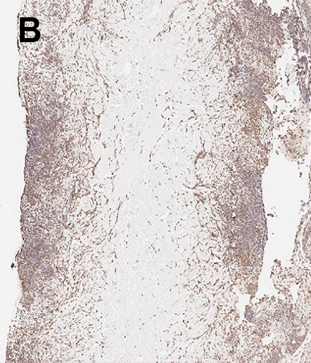
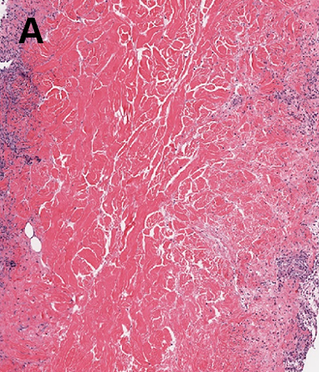
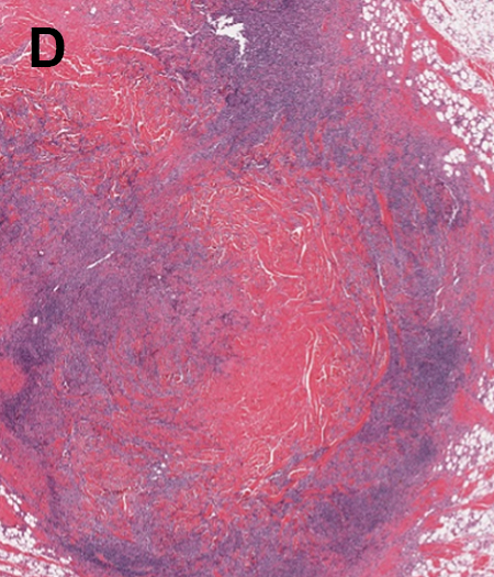
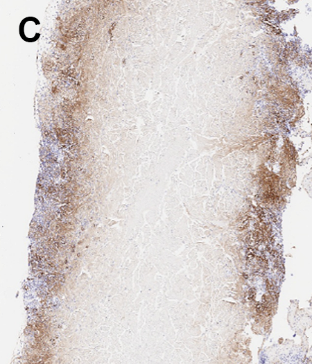
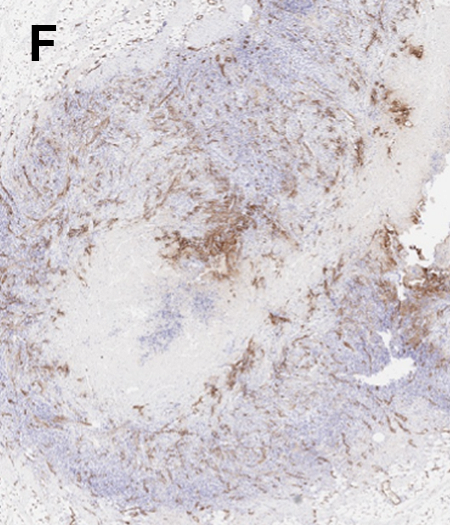
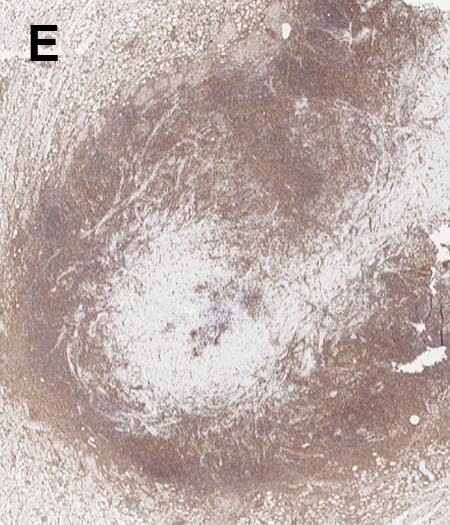
Figure 1: Histological images of explanted grafts after one week following surgery: (A-C) H&E, vimentin and Factor VIII staining for control ADM grafts and (D-F) H&E, vimentin and Factor VIII staining for MS ADM grafts. Cells are represented by purple dots in H&E staining and brown dots in vimentin and Factor VIII staining.
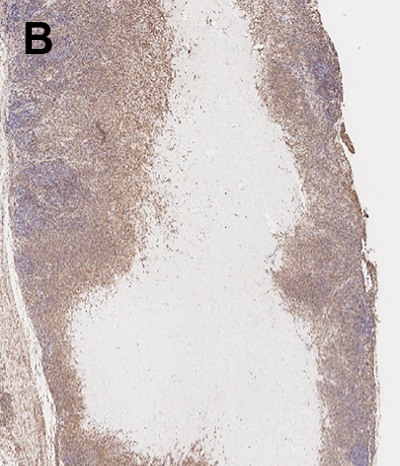
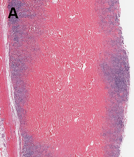
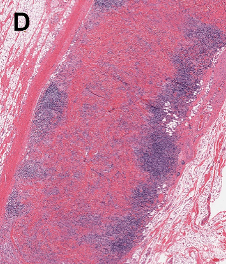
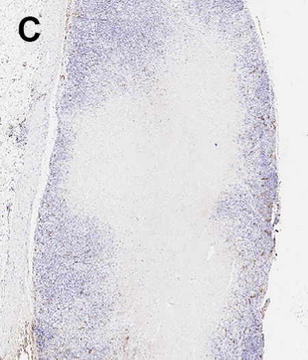
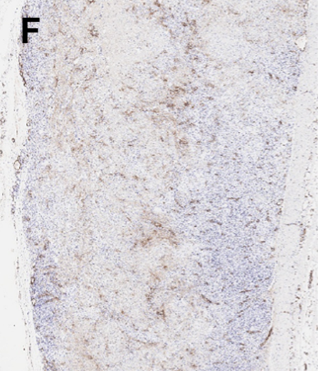
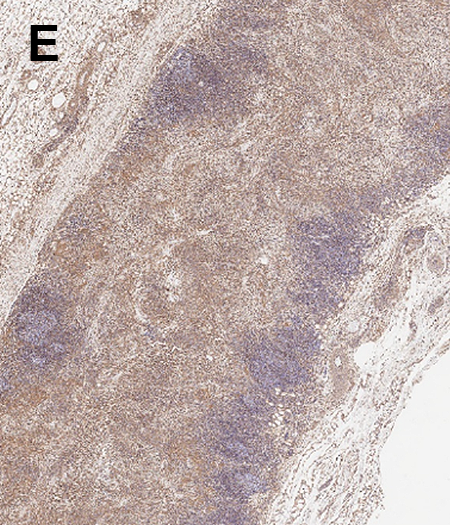
Figure 2: Histological images of explanted grafts after two weeks following surgery: (A-C) H&E, vimentin and Factor VIII staining for control ADM grafts and (D-F) H&E, vimentin and Factor VIII staining for MS ADM grafts.
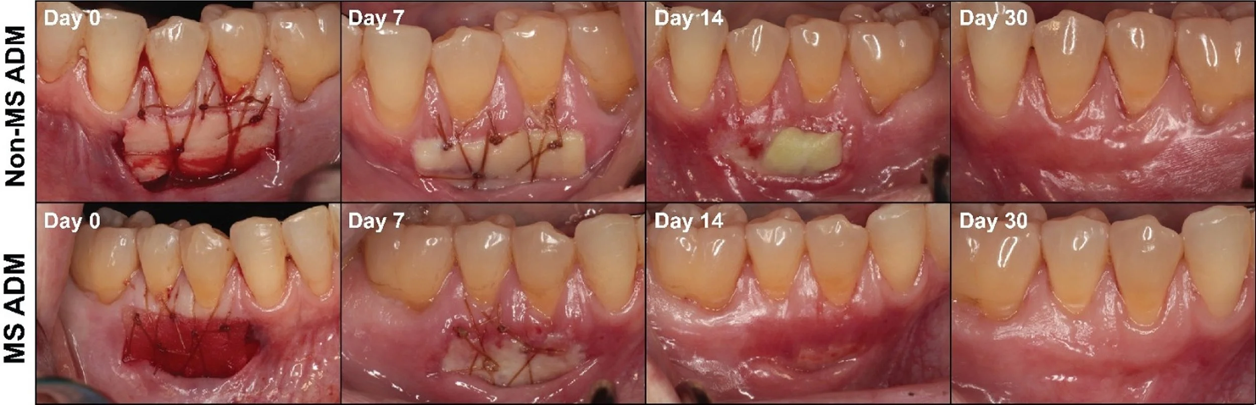
Figure 3: Representative images demonstrating comparative restoration of gingiva within the same subject for non-MS ADM (top row) and MS ADM (bottom row) after graft placement over 30 days.
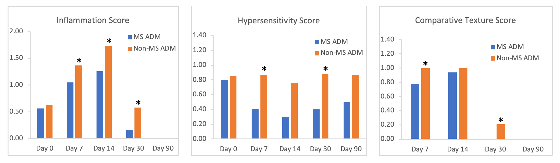
Figure 4: (A) Inflammation score; (B) hypersensitivity score; and (C) comparative texture score. Asterisks denote statistical significance (p<0.05)
Fast Incorporation
Early clinical applications of MS ADM grafts included usage for soft tissue augmentation in periodontology. A prospective, intra-patient clinical study was conducted in which MS ADM was compared to a market-leading, commercially available for gum grafting in 10 patients with gingival recession⁵. Here, one graft of each type was placed in sites with <1mm of attached gingiva located within the same patient.
Following graft placement, rapid incorporation was observed for MS ADM grafts with significant visually observable differences compared to non-microsurfaced ADM (non-MS ADM) over the first two weeks post-surgery (Figure 3). In particular, more uniform blood penetration was noticed immediately after implantation, and substantially less original graft material was seen at 1 and 2 weeks. Furthermore, blinded observers noted that 100% of the MS ADM graft sites exhibited more graft remodeling than those treated with non-MS ADM at 7 days and 90% at 14 days. By 30 days, application of MS ADM grafts resulted in successful restoration of >1mm of keratinized gingiva. There were also two displaced grafts in the non-MS ADM group compared to zero in the MS ADM group.
Striking differences in multiple outcome measures including inflammation, hypersensitivity and comparative texture were also noted during a period of 30 days after application (Figure 4). More specifically, scores for inflammation and hypersensitivity were significantly less in the MS ADM group while comparative texture was significantly improved compared to the non-MS ADM group. The differences were most pronounced at Day 7 and Day 14 suggesting that the partial thickness micro-cuts on MS ADM grafts result in a more rapid repair response. As anticipated, non-MS ADM grafts still support tissue regeneration over an extended period of time, but they achieved this response at a slower pace.
¹ Wu Y, Cheng G, Rotenberg S. (2025). “Root coverage using a microsurfaced acellular dermal matrix: a retrospective case series.” Clinical Advances in Periodontics. doi: 10.1102/cap.10361.
² Tao CY. (2025). “Evaluating the efficacy of micro-cut design allografts in treating multiple gingival recession defects: a case series.” Presented at the EuroPerio Congress Meeting; Vienna, Austria.
³ Data on file at CellTherX™.
⁴ Marinelli LM, Krauland K, Alcorta S, Homsombath B, Fagan S, Craft-Coffman B, Hassan Z. (2023). “Histological evaluation of wound-bed preparedness following microsurfaced skin grafts for the treatment of deep burn wounds: results from a randomized controlled trial.” Presented at the AMSUS Meeting; National Harbor, MD.
⁵ McGuire MK, Scheyer ET, Davis KE, Gunsolley JC, Heard RH. (2026). “Use of micro-surfaced acellular dermal matrix for the generation of site appropriate keratinized tissue in soft tissue augmentation healing: Proof of concept.” Clin Adv Periodontics. doi: 10.1002/cap.70073.